Neurovascular Coupling of Cortical Inhibitory Neurons
We are investigating neurovascular coupling and the contribution of excitatory and inhibitory neurons to blood flow regulation in the rodent cortex.
Personnel
- Alberto Vazquez (Principal Investigator)
- Alain Altamirano-Espinoza
Project Summary
Increases in neural activity produce similar increases in regional cerebral blood flow (CBF), cerebral blood volume (CBV) and the cerebral metabolic rate of oxygen consumption (CMRO2). These physiological responses give rise the blood oxygenation level dependent (BOLD) effect that is routinely used in functional MRI studies of brain function in animals and humans.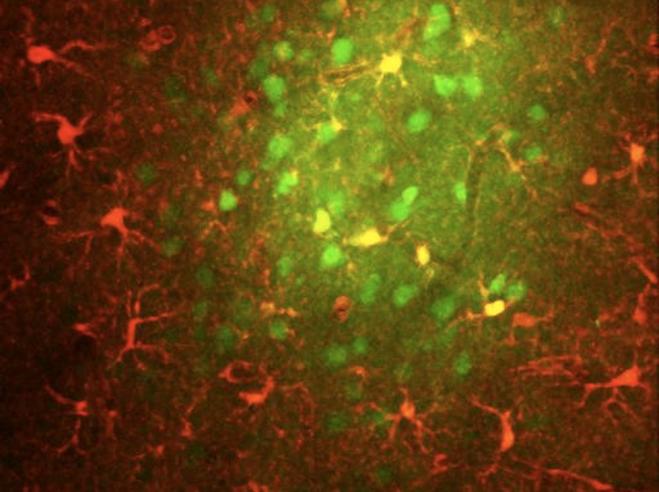 |
The brain is composed of a very diverse population of cells that have distinct contributions to network activity and also to vascular function and metabolic load. The goal of this project is to use optogenetic actuators (and silencers) to determine the contributions of specific cell types along the neurovascular signaling pathway to vascular responses. This project is guided by the following general questions: (a) Do changes in excitatory neuronal activity alone modulate proximal arterial vascular tone? Is this mechanism specific to prostaglandin signaling? (b) Do changes in inhibitory neuronal activity modulate proximal arterial vascular tone? Is this mechanism specific to nitrous oxide release? (c) Do changes in astrocyte calcium modulate arterial vascular tone? Is glutamate release necessary? (d) Can the dilation of vascular smooth muscle modulate neural activity? This project is relevant to studies of human brain function in health and disease because dysfunction of distinct brain cell types has been implicated with specific neurological disorders. |
| We have targeted different populations of inhibitory neurons using transgenic mice expressing cre recombinase in neuronal populations of interest and performed experiments in awake head-fixed mice. Channelrhodopsin-2 (ChR2) was virally transduced in the somato-sensory cortex of these mice and hemodynamic responses sensitive to blood flow were recorded during optogenetic and whisker stimulation. NOS expressing neurons are not as numerous and some of these have long-projecting processes. |
Below is a summary of the changes in blood flow evoked by optogenetic and whisker stimulation for 1-sec (top row) and 4-sec (bottom row) while modulating the optogenetic pulse duration from 2ms to 30ms to evoke different amounts of activity. Whisker stimulation is shows in black.
Below are the changes we observed when evoked optogenetically stimulating these different mice at different frequencies for 1-sec (top row) and 4-sec (bottom row).