Chemical Exchange-Sensitive MRI
The chemical exchange (CE) effect has recently been exploited as a powerful method that is highly sensitive to detecting biological chemicals present in low concentrations. Measurement of the CE effect is possible due to the exchange of protons between specific chemicals and water, which results in a change in the detectable water signal. The detection of endogenous biochemical agents, such as metabolites, peptides and proteins, by CE-derived image contrast is particularly attractive because it can be used to probe the tissue microenvironment, which includes tissue pH, temperature and concentration of agents with exchangeable protons. In our laboratory, we have developed novel CE MRI approaches that can be specifically tuned to amide, amine and hydroxyl protons in proteins, peptides and metabolites, and is currently being applied to detect changes in the tissue microenvironment present in disease models. In this way, the development of CE MRI is important for early detection of disease and to determine the effectiveness and progress of treatments.
Personnel
- Tao Jin (Principal Investigator)
- Bistra Iordanova
Project Summary
Imaging of tissue intracellular pH changes
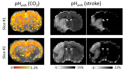 |
We have developed a few endogenous MRI techniques (APEX, APT*, and pHenh) which are sensitive to intracellular pH. For example, pHenh has high pH-sensitivity and can detect the tissue acidosis due to hypercapnic challenge or in acute ischemic stroke. These techniques can be used for the evaluation of tissue metabolic status in diseases such as stroke and TBI. |
Imaging of glucose uptake with non-labeled glucose or glucose analogs
| We have developed a glucoCESL technique to detect the glucose uptake and metabolism with an injection of unlabeled glucose or glucose analogs such as 2-deoxy-glucose and 3-O-methyl-glucose. In brain tumor, this approach can detect a BBB leakage in the initial period, and is sensitive to the glucose uptake in the later period. This technique can also be used for the study of stroke, TBI, and neurodegeneration. | 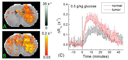 |