Olfactory Bulb Imaging
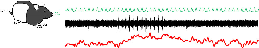
We are investigating functional hyperemia to evoked neural activity and its direct relevance to changes in the fMRI signal. We chose the rodent olfactory bulb as our model system because of its well known neurophysiology that allows us to examine hemodynamic responses to specific neuronal events.
Personnel
- Hiro Fukuda (Principal Investigator)
- Alex Poplawsky
Project Summary
The olfactory bulb has a laminarized structure, whereby each layer contains distinctive cell types and synapses, and receives specific input fibers. This makes the olfactory bulb ideal for studying the neural basis of fMRI. The goal of this project is to establish detailed relationships of the hemodynamic responses to the activity of specific cell types in the individual olfactory bulb layers and how cortical feedback and neuromodulators affect such relationships, which can serve as a reference for neurovascular coupling in many diseases expressing olfactory dysfunction.
We have recently demonstrated that 1) hemodynamic responses are regulated at a laminar level and 2) synaptically activated inhibitory neurons following stimulation of the lateral olfactory tract (LOT) and anterior commissure (AC) increased hemodynamic response. However, several questions remain unanswered: 1) What is the major mediators of fMRI responses evoked by inhibitory neurons: nitrous oxide, neuropeptides, GABA or something else? 2) Does astrocytic activity (e.g., glutamate or GABA reuptake) play a key role in the fMRI response evoked by inhibitory neurons? 3) What makes the time courses of fMRI responses evoked by LOT and AC stimulation so different despite the activation of the same inhibitory neurons? 4) What causes the negative fMRI responses in the side contralateral to the LOT stimulation? 5) Can activity of inhibitory neurons during sensory processing also produce measurable fMRI response? 6) How do neuromodulatory and cortical feedback inputs affect the odor stimulation-evoked layer-specific fMRI response? 7) How do neural plastic changes modulate layer-specific fMRI response?
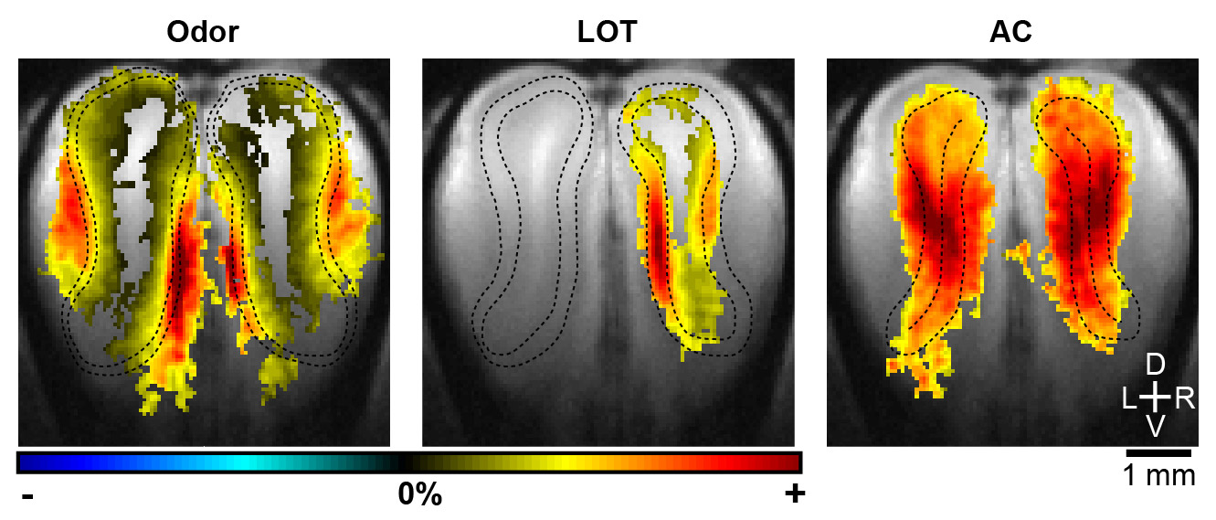 |
| Adapted from Poplawsky AJ, et al. (in press) |
| The laminar cerebral blood volume-weighted fMRI responses to odor (5% amyl acetate in mineral oil) and electrical stimulations of LOT and AC stimulations, respectively. Group activation maps: n=8 rats, threshold of p<0.025 for voxel- and family-wise error correction (i.e., 30 voxel cluster minimum), black dashed lines surround the layer of preferentially evoked synaptic activity determined in the T2-weighted anatomical image (i.e., the grayscale underlay). Colored scale bars: ±4% for AC; ±7% for LOT; ±10% for odor. |